
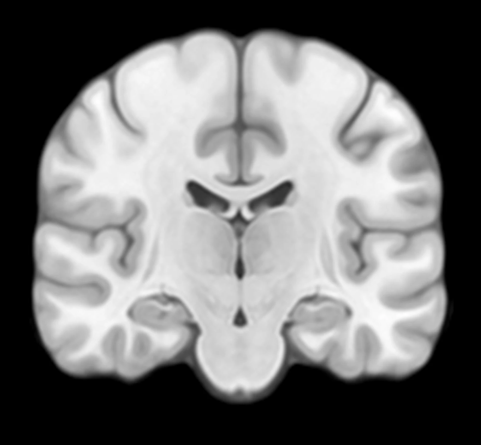
1Measures the regions that matter
The regions affected earliest are measured automatically from a standard MRI. No manual tracing.
Meet CeDawnMeet CeDawn · MRI, measured
See what matters. Earlier. Cerebramha.
RLCOR · y −18.6 mmT1 · 1 mm hippocampus ventriclesnormal range for age and sexthis measurement
36-month MCI to AD
58%High
Model: imaging + clinical. Inputs: MMSE 24 · APOE4 ε4 ×1. Drivers: hippocampus, entorhinal cortex, ventricles.
The problem
Alzheimer's is most treatable early, and the newest therapies only work at that stage. The signal is already in the MRI, in shrinkage too subtle to quantify by eye.
RLCOR · y −18.6 mmT1 · 1 mmHippocampus. Smaller than it should be? Hard to say.
Read visually, slice by slice. No number.
RLCOR · y −18.6 mmT1 · 1 mm hippocampusHippocampus, 2.74 ml. Normal: 3.40 to 3.92.
6th percentile, z -1.55, for her age and sex.
Hippocampus
Measured against normal
2.74 mlnormal 3.40–3.92
How it works
Three steps from a standard MRI to a one-page report.
The regions affected earliest are measured automatically from a standard MRI. No manual tracing.
normal range for age and sexthis measurement
Each region is placed against a reference population matched for age and sex: the measurement, the normal range, and the percentile.
36-month estimate
58%High
A 36-month estimate of progression from MCI to Alzheimer's, from the scan, age, and sex. Cognitive or genetic data refine it.
Why it's different
A score with no reasoning is unusable to a clinician. CeDawn shows the measurement, the normal range, and the regions behind the estimate.
RLCOR · y −18.6 mmT1 · 1 mm hippocampus ventriclesWhere we are
CeDawn is a research platform. It runs end to end today, and we're validating it with clinical and research partners. It is not a cleared medical device and does not replace clinical judgment. We're building the evidence base for clearance, with research groups like yours.
Runs end to end todayValidating with clinical partnersNot a cleared medical device
Building the validation
Run CeDawn on your cohorts. You get a novel quantitative measure and progression estimate for your own work, with co-authorship on what comes out of it. Collaborators, not vendors.
Research groups · co-authorship